InFocus™ Dynamic Optical Focusing Systems
Dynamic Optical Focusing and Spherical Correction
If your microscope doesn’t have InFocus…
it may not be in focus.
- Imparts DYNAMIC OPTICAL FOCUSING to Almost ANY Infinity-Corrected Microscope
- Corrects Spherical Aberration Better than Correction Collared Objectives
- Works with ALL Infinity-Corrected Objectives
- Color-matched for Zeiss, Leica, Nikon or Olympus—and others
- Allows YOU to Test Your Objective’s Performance
- The Better The Objective You Use—the More You Need InFocus
- Breadboard or Stand Options

InFocus Series
• Magnification Tables
• Instruction Manual
NEW
The InFocus PRO is the same as the regular InFocus, but has provision for interchangeable front lens options. This means that by using the non-standard LENS 2, the full range of PLUS can be utilized. LENS 1 retains the standard “near-balanced” range of the regular InFocus.
Previously, the Plus-Weighted C-mount served this purpose—but only with C-mount cameras or sensors. Now, the PLUS range can be utilized for all formats. In addition, the Plus-Weighted C-mount can be used with the InFocus PRO to even greater PLUS potentials than before.
If your work requires deep cell translations or involves mismatched media, the InFocus PRO may better suit your needs. In all other respects, its use is the same. As with the regular InFocus, the PRO model is available for use with infinity-corrected objectives made by most microscope objective manufacturers.
InFocus makes the difference…
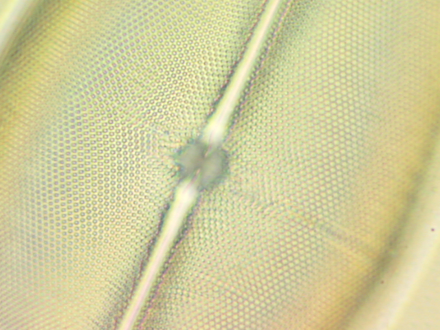
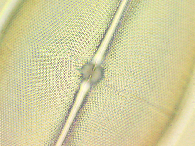
Diatom: Pleurosigma in Pleurax (RI 1.8) without InFocus (L) and with InFocus®
InFocus is a unique optical device—in fact, a system—that allows you to realize the full potentials of optical microscopy. InFocus can be thought of as the “ultimate” InfiniTube when infinity-corrected objectives are used with it. But where the simpler InfiniTubes impart minor focus adjustment, InFocus imparts true optical focusing. Whether you use InFocus as a self-contained microscope in a breadboard setup—or attached to a microscope stand—it provides added degrees of convenience and perfection. True optical focusing means that a microscope equipped with InFocus can be deliberately defocused so that a new position can be set for the relative working distance of microscope object to objective. This is more significant than may at first be thought.
Beyond its simpler industrial and general uses, InFocus is a significant breakthrough wherever cover glasses must be used. In biological and high-tech industrial applications, this means that it can be used to diminish or correct the problem of preparation induced spherical aberration (SA). (In SA, an optical system cannot focus all the light at a common plane resulting in a fogging of image details). This is the single most overlooked reason for poor microscope imagery. Even the finest optics can be thrown out of correction when cover glasses, thick or highly-refractive media are used.
This is all due to the fact that the relative position of the objective’s focus is determined by the thickness of the cover glass (or lack thereof) and the medium in which the subject is immersed. Manufacturers claim to correct their biological or special industrial objectives for use with a defined cover glass thickness with the preparation just beneath it. Deviating from this factory setting creates (for various optical reasons) SA whereby image detail is never properly focused or captured. The resulting image can even become useless blurring—particularly in fluorescence applications. But, unless a perfect objective is used perfectly with a cover glass (biologically, usually set for 0.17mm) designed for it—with a specimen mounted just beneath in a mounting medium of specific refractive index—SA and a degraded image—are inevitable. This is why most photomicrographs are imperfect. As a consequence many published images are more or less “fuzzy” compared to how they would be if captured with a spherically corrected system.
Human Bone Marrow. Olympus UIS-2 60x/0.90NA Plan Fluorite Objective.
1x Factor Direct to Sensor. Without InFocus and with InFocus.
Taken by H Jay Margolis with his original improved InFocus prototype.
DYNAMIC FOCUSING: SUPERIOR TO CORRECTION COLLARS

By 1830, both Amici and Lister had independently observed that spherical aberration was induced by using cover glasses with high apertured microscope objectives. This severely diminished image quality.
In 1837, Andrew Ross—acting on Lister’s suggestion—made the first objective with a built-in correction collar. The correction collar has remained the only means to compensate preparation-induced aberrations ever since.
With correction collars, the objective’s own lens system can be actively separated. At a normal null position (with biological objectives, generally 0.17mm cover/medium thickness), a correctly made preparation will provide a “best image.” If the cover/medium thickness deviates from the norm, the correction collar is turned to set a new “best image” which is refocused by using the mechanical focus. Turning the correction collar itself does not impart focal translation. Consequently, correction collars activate optical changes within the objective and only for that particular objective. When used with infinity-corrected systems, correction collared objectives operate on the beam characteristics before they are acted upon by the infinity tube lens system. The infinity tube lens system remains STATIC.
With InFocus, one common module—usable with virtually every objective—acts almost precisely in reverse. With InFocus, the objective remains STATIC but the infinity tube lens system becomes DYNAMIC. In effect, InFocus “forces” an objective to seek a new frontal focus. Then, the previous plane can be re-established by the mechanical focus with increased fidelity because InFocus has automatically compensated for all factors: the cover glass’ thickness, deviations in media refractive indices and refractive errors. Because InFocus imparts DYNAMIC OPTICAL FOCUSING, different depths can be focused upon (once the initial “best image” is obtained). This feature is of immense advantage to those who cannot allow the objective to move in any way, particularly when observing delicate tissues which are easily displaced by compression or when industrial processes can come in contact with the objective if it is moved to focus.
Since InFocus is a module rather than a feature built into a single objective, it can be used to critically test the quality of objectives. If the chosen objective is claimed to be factory-set for 0.17mm and the InFocus cannot correct its image, the objective is probably faulty. And, since the InFocus works with all objectives, you need not buy correction collared objectives if a simpler mounting is available. If all this were not enough, InFocus can be mounted on dovetails so that one, usable with one make of objective and be switched with another InFocus usable with another make. You can actually choose which objective to use from among various makes and types—a dream since the early years of microscopy.
Background of InFocus. Until InFocus, there were two ways to correct SA in microscopes. One was to push in or pull out the drawtube of old-fashioned monocular microscopes. For every 10mm of movement, approximately 0.01mm of cover glass correction could be made. This was not very efficient, especially with thicker preparations where only about 20mm of down movement was even possible. The other way was to use microscope objectives fitted with correction collars that separated the lens elements and reset the focus obtained. But this method—still used today—is cumbersome, difficult and usable only with objectives so-equipped to begin with. Objectives which do not incorporate correction collars can never be utilized to advantage. More, just by touching the objective to make the correction, decentering or compression of the sample are almost assured.
The original InFocus eventually went on to be motorized and computer-activated in order to determine its best-contrast setting, pricing itself into a small, exclusive market. Although simpler systems were offered, their functional ranges needed supplemental lenses to extend operational efficiency where needed. Still, the original InFocus was such an advance, that it went on to win two R&D 100 awards.
In recent years, Infinity Photo-Optical introduced the K-Series long-distance microscopes equipped with patented CentriTel® focusing. CentriTel is an internal focusing system which does so without essential magnification change. This allowed us to revisit the InFocus concept and apply features derived from CentriTel to it. During that time, camera sensor technology advanced considerably. As a result, it is now possible to offer an improved InFocus Module that is simpler, more universal—and does not require computer-activation in order to determine its best setting—putting the price and image enhancements of InFocus within reach of most research and industrial microscopists. In addition, the improved InFocus has minimal magnification change through its focal translation and centers on a c.1x factor.
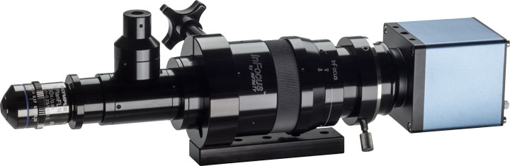
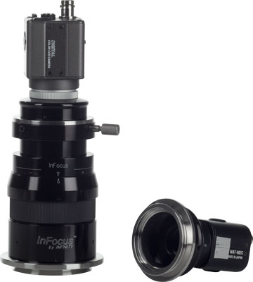
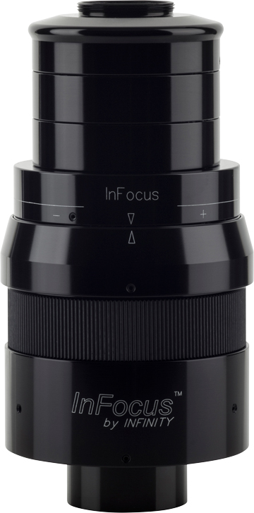
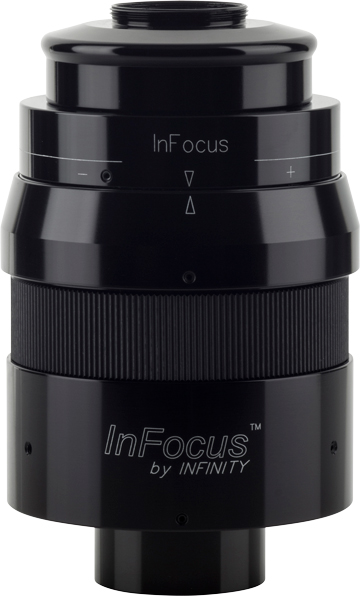
What InFocus is. InFocus is an integrated microscope tube with patented optics, complete with a tube lens for use with infinity-corrected objectives of various makes (more on this later). When mated to an infinity-corrected microscope objective (either integrated in a breadboard setup or on a microscope stand), InFocus is a complete microscope—but with one very considerable difference: it has an internal focusing adjustment which is actually finer than any mechanical focus can be: A dynamic optical focusing system. Turning a knurled ring activates InFocus to translate focus from above, through and below the object. For industrial setups, InFocus allows focal translation and “touch up” even if the unit is bolted in place. For biological or high-tech industrial applications, it goes one step further by utilizing its ability to alter tube length—as a drawtube would—but without changing outer dimensions. Also, its operative range is far, far greater than that afforded by any drawtube. As a result, InFocus imparts the ability to correct spherical aberration to any objective used with it—even those already equipped with correction collars (which should be then be set at their null position).
By adding InFocus’ dynamic optical focusing to any microscope—either onto the stand or in a breadboard system—it becomes possible to do things that previously required special equipment. Now, every microscope is micromanipulator-ready because focus can be adjusted without stage movement. Now, every microscope can be equipped beyond the capabilities of what correction collars—which do NOT provide dynamic optical focusing—can do. In fact, the degree of correction possible from InFocus is considerably greater than that of typical correction collar-equipped objectives. And, if quality results cannot be obtained at a reasonable setting of the InFocus, your objective may be a defective one.
The improved InFocus has an operating range approximately four times that of the original. This opens new vistas for use. Now, it is possible to set the initial SA correct position and, from that starting point, use the optical focus instead of the mechanical to select various Z-depth planes. There are now two ways to use the InFocus for specific results. The first is to set it for SA correction. For single one-plane image capture, that is sufficient. But for Z-stacking (with software such as Helicon Focus™ or Zarene™, the SA correction can be made and then re-established at another depth obtained by using the mechanical focus. That is the traditional way to use InFocus. However, it is now possible—due to the extended range of the improved module—to set SA correction and then use only the InFocus to dynamically select different depths—abandoning the use of mechanical focus altogether. This feature of the improved module makes it possible to focus without moving the objective through delicate membranes which might otherwise be subject to medium compression. What is centered to start is still centered at the finish of a focal sweep.
Regardless of the method used, a microscope equipped with InFocus can provide images better than one which is not. It is as simple as that. But it was neither simple nor easy to make that happen during the development of InFocus.

Four Major Brands—THREE solutions required. Years ago, it was reasonably simple to use one brand of microscope objective next to another. Makers pledged to use the “RMS Thread” to fit objectives to microscopes and, even if one make used a slightly different tube length, the drawtube could correct that. Most all microscopes were monoculars. Even if one maker’s objective needed a different eyepiece to work with it than another’s, it was easy to pull one eyepiece out and put another in its place. But with modern binocular microscopes and the advent of flat field (plan) objectives, things got complex. Then, when the advantages of infinity-correction became too obvious to ignore, makers settled upon their own optical solutions. Zeiss chose to keep 160mm tube length and RMS mounting thread (now also offering M27 x 0.75), the same objective length as before and transferred their compensation from “KPL” eyepieces to the infinity tube lens. Leica did similarly, selecting 200mm as the tube length and putting their “HC/Delta” correction in the tube lens. Thus, Zeiss and Leica use “color-specific” tube lenses. Nikon and Olympus chose to use various threads and dimensions (Nikon 200mm and Olympus 180mm tubelength), etc. but achieve all color correction in the objective itself. Nikon and Olympus tube lenses can therefore be termed “neutral.” Others also use the “neutral” correction, such as Mitutoyo, Edmund and Infinity Photo-Optical.
In order to offer InFocus as a viable product, it was necessary to replicate the corrections for the two “color-specific” brands and provide a “neutral” system for all others. Consequently, there are not one—but three InFocus modules. The Zeiss and Nikon/Olympus modules are the same overall length while the Leica is 9.5mm longer. The tube focal length has been selected to be c.160mm; therefore, a Zeiss objective operates at its rated magnification, an Olympus at 0.88x its rated magnification, and a Nikon, Mitutoyo or Infinity Photo-Optical objective at 0.80x its rated magnification. However, amplifiers can attach to the InFocus to permit alteration of these “raw” factors. This approach allowed us to set the rear accessory distances to be the same.
With the successful development of three different InFocus modules—all compatible with a whole line of accessories, the InFocus is properly a system unto itself. Bluntly, if your microscope is not equipped with InFocus, it probably is not in focus.
InFocus is easy to use. Basically, all you need to do is to:
- Mount the InFocus via its proper dovetail adapter on a microscope stand or, if used in a breadboard setup, use a T-tube to secure it. Clamps and other accessories permit this to be done (see Drawings). If the bottom part of the InFocus contacts any part of your microscope, a small T-tube should be interfaced—but this is unlikely on microscope stands.
- Common parfocalize the system. You can now absolutely parfocal your microscope. When your microscope is set for the common parfocal position, swinging one objective into place from another results not in a generally focused image, but one which is essentially at the same plane of focus as the other.
- To SET for common parfocality, use a low power (e.g., 10x) objective and then use the highest numerically apertured high dry objective (HDO). Generally, this should be a 40/0.75 or 60/0.90 objective. Focus the 10x and then swing to the HDO. Swing back to the 10x and turn InFocus’ adjustment to reset focus. Repeat this. After the 10x has been refocused by the InFocus a second time, switching between the objectives will result in the same plane being in focus, regardless which objective is used. The microscope is now set for common parfocality. The goal is to have your microscope set so that “infinity” is virtually absolute between two objectives whose depth of field v. depth of focus characteristics are near opposite. This establishes the starting point for InFocus’ operation.
- Your InFocus was set at the factory so that when the two arrows on its focusing ring/body face each other, it is at infinity. If you wish to reset them, use a 2-56 Allen wrench (2mm metric) to slightly detent the metal colored set screw under the arrow, turn, and gently lock in place.
- You will note MINUS (-) and PLUS (+) markings on the InFocus body. Minus makes the unit correct for progressively thinner preparations and Plus makes the unit correct for progressively thicker preparations. Please see the InFocus Graphics for an explanation as to what these mean exactly. In any case, the InFocus can be used as simply as capturing one image, then another, all while moving the InFocus control in several steps and re-establishing the same plane by mechanical means. InFocus CAN be as simple to use as that: One image will stand out as the best; one image will be the one you wish to “publish.”
Advanced Use. While indiscriminate use of InFocus is possible—as outlined above—InFocus comes into its own when it is used with more technical attention. The improved InFocus now permits two techniques to be utilized:
- Mechanical Focus Reset (MFR).
- Dynamic Optical Focus Sweep (DOFS).
Mechanical Focus Reset. Here, the InFocus is progressively turned and a mechanical focus re-established for the plane of interest. The contrast will either improve or deteriorate depending on the match for SA correction. If a thin preparation is used, first start with the Minus side of the translation. If a thick preparation is used, “weight” the translation to the Plus. Observe the increase in contrast at each setting. If you are using InFocus with a camera that allows electronic finder magnification increase, this may be very simple to determine. You can focus at different Z-depths and process for stacking or deconvolution, later.
Dynamic Optical Focus Sweep. This technique is possible due to the increased efficiency of the improved InFocus module. The best contrast is found by using MFR. It differs thereafter, since, from that position, the InFocus is used to seek different Z-depths. The mechanical focus is not used at all.
[The improved InFocus has such a sufficient operating range, that even a diatom mounted in a medium such as Pleurax (1.8 index of refraction) can be brought to greatest contrast at c.90-degrees of the turn to Plus, leaving at least that many degrees of additional turn of the InFocus dial. Even then, if the InFocus is turned still more to the Plus, the image will degrade. In effect, the InFocus is then “asking” for a diatom at an even lower level to be imaged.]DOFS is particularly useful where delicate membranes must be imaged and where any movement of objective could cause displacement, rendering the collective images captured useless. Since any focal changes from the InFocus are independent of connection or contact with the objective and preparation, it allows images to be captured as never before.
InFocus Graphics. Full details on how the InFocus works, what preparations it can be used with and the SA corrections possible are contained in the InFocus Graphics section attached to the Instructions PDF. The InFocus Graphics also show the relative positions of the objective in different media and/or thickness situations.
Applications and Camera Formats. If you are doing deep cell translations; attempting point-spread functions; 3-D imaging or confocal microscopy; if you are examining wafers and wish to avoid all possible contact; if you are building a breadboard instrument—or simply trying to capture accurate imagery—InFocus will allow you to do things previously thought impossible. Every microscope now becomes micromanipulator-ready. And, InFocus has a whole system of accessories to back it. With them, it is possible to use virtually every type of camera that has ever been used in photomicrography. This holds especially true if you have one of the advanced research-grade C-mount cameras from the major microscope makers. All their software can be utilized.
Modern digital cameras are now so efficient that the improvement in contrast provided by InFocus is easy to see. This is especially true when electronic magnification is increased on the display or to a monitor. The determination of the point where contrast is maximized by InFocus is such, that computer determination (which was once considered a necessity) is no longer required.
The Plus Weighting C-Mount is ideal for setting the Plus range for deep translations or high-index media.
The Plus-Weighting C-Mount. Most research-grade cameras offered by the major microscope makers are 2/3-in. C-mount types. If deep focal translations, high refractive index media, thick covers, etc., are used or contemplated, the Plus-range of the InFocus becomes more and more necessary to achieve corrections. The Plus-Weighting C-mount can be shortened in two steps of 12mm so that common parfocality occurs with most of the Plus-range still able to be used. If you are planning to do such work, we highly recommend the Plus-Weighting C-mount instead of the regular types we offer.
In Sum. InFocus is not for everyone. Not everyone will need to publish the finest imagery. Not every industrial process needs to be monitored from a fixed position. Not every biological sample needs to be recorded. But, if you think InFocus will assist your research or high-tech application, contact us. We will let you know how to capture imagery in focus by InFocus.